BLOG
GRAM STAINING TECHNIQUE – PHƯƠNG PHÁP NHUỘM MÀU CHO VI KHUẨN
When you stain bacteria with the Gram staining technique, then Gram Gram-positive bacteria are stained in “a blue color,” and Gram Gram-negative bacteria are in “Red color.” why do they have different colors? The real understanding is the different cell wall structures.
It was developed by a Danish physician in 1884; what is the basic principle?
What is step number one?
Let’s suppose you have “sputum of patient. What is your first step if you want to stain the sputum and find gram-positive bacteria present, Gram-negative bacteria present, or both bacteria present?
The first step is to take a sample and apply the sample on the “slide.”
The first thing you do in the laboratory after applying the “sputum” or the sample on the slide is heat fix. You heat the “slide” a little bit. So, the bacteria will become fixated on the slides.
It should not happen that you dip the slide into different types of colors and have bacteria wash away.
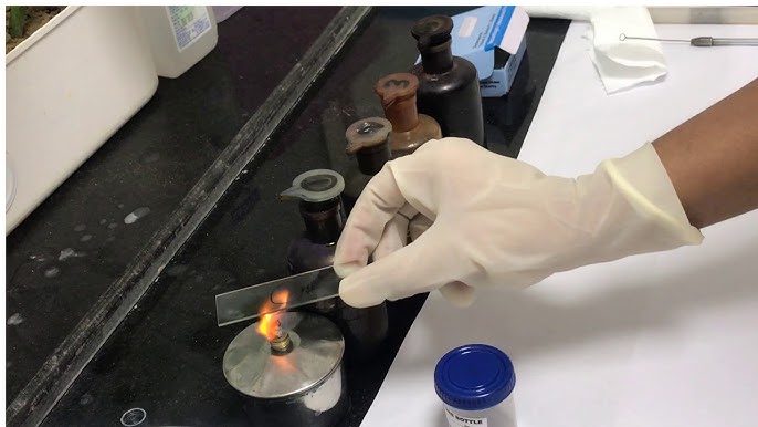
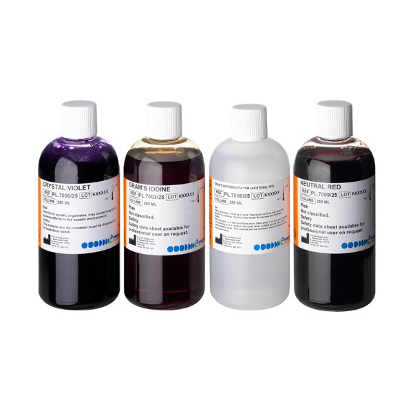
The next step is to color the slide with “crystal violet dye stain.” for example, if you have a container that contains “crystal violet dye stain,” we stain it for 60 seconds with crystal violet. Then, bring it out and wash it with water. What will the color of these bacteria be?
Let’s suppose these are gram-positive and these are gram-negative
Now, all of them have a “violet color or blue color.” The question is, why are all of them stained in blue?
The answer is the crystal violet dye can go into the peptidoglycan layer and bind with the Peptidoglycan of both types of bacteria because “crystal violet dye” can easily go into the porins of the Gram negative Bacteria and bind with Peptidoglycan.
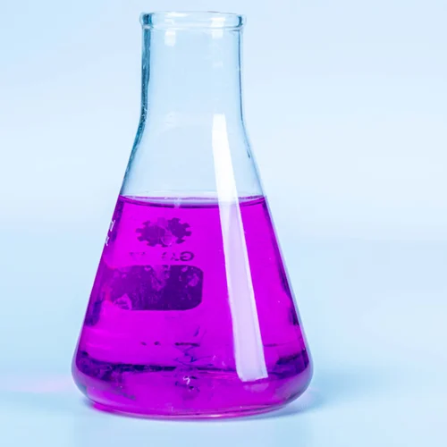
The next step is to have “iodine as a mordant.” now, the slide will go into the container with “iodine” in 60 seconds. When it is stained with iodine, the iodine molecule will make a complex with the crystal violet; after this, we wash/rinse it with water; what will be the color? Now, in this step, all of them are still looking “blue.”
Now, you should think about why iodine is added.
Let’s remember this: because Gram Gram-positive bacteria have big Peptidoglycan, they should take much violet; the cell wall of the Gram-negative bacteria is thin, which is why they take less violet.
If we don’t add iodine, what will happen? Then, both of them will be strongly stained with crystal violet. Later on, with alcohol, we cannot wash crystal violet out of gram-negative; what is iodine doing? Iodine will make the cross-link in the molecule of violet; when iodine binds with crystal violet, then they can easily washed by alcohol out of gram-negative bacteria.
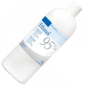
Next step: is alcohol ethanol 95%? Of course, you bring the slide out and rinse with water. Add it to alcohol 95% ethanol or acetone alcohol solution and bring it out. Actually, alcohol will dissolve/disrupt the outer membrane of gram-negative bacteria, and of course, gram-positive bacteria do not have an outer membrane.
This is the point you must understand
Gram-negative bacteria have little Peptidoglycan with little cross-linked, and opposite to that, gram-positive bacteria have a huge quantity of Peptidoglycan. What happens? When you dip this slide into alcohol gram-negative material, it loses its outer membrane, and Peptidoglycan is so thin and so widely porous that the iodine crystal violet complex falls out of this Peptidoglycan; in this way, Gram-negative bacteria becomes decolorized. In this step, Gram-negative bacteria lost their “blue color.” Unlike gram-negative bacteria, gram-positive bacteria still remain blue.
If you have not added iodine, the color will remain sticky on both.
Next step: we all slide into another dye that is “safranin” It is red in color, and bring it out, wash it, then dry it with “blotting paper” Then, it is ready to be observed under the light microscope, then Gram-Positive Bacteria remain blue, purple color, and gram-negative bacteria after washing alcohol crystal violet and iodine complex has washed away from negative bacteria then now safranin will be added to the gram-negative bacteria will turn into red color.
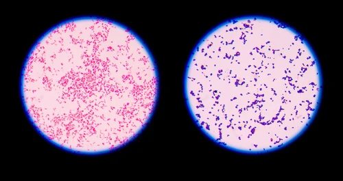
This is a gram staining technique. After this technique, you can learn what type of bacteria is present and its shape.
Huỳnh Trụ


